The human brain is, contrary to popular belief, not at all grey but pinkish. When preserved in formalin a dead brain will however turn grey, and this is likely why the brain so often has been portrayed as grey in popular culture.
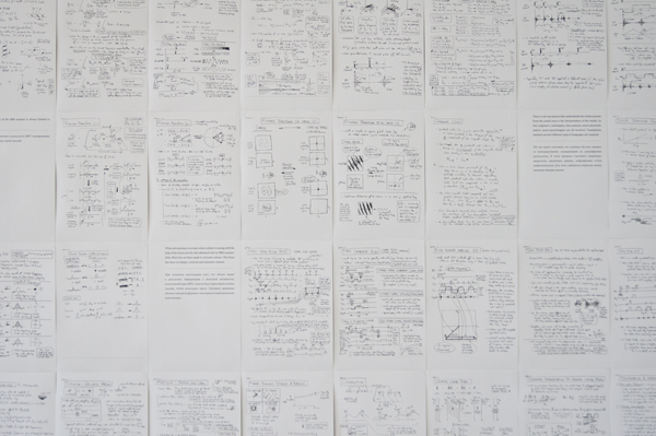
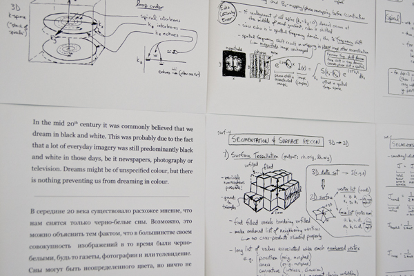
In contrast, MRI images are all in greyscale. The data retrieved during a brain scan is first organized in the so called K-space, a greyscale raster containing all the coordinates of the collected data and from this a 2 dimensional black and white image is constructed.
A finished MRI image might be colourized to enhance clarity or to make for a more spectacular presentation, but these colours are not directly based on information collected during the MRI scan, and are subjected to a fair amount of speculation. Depending on what a scientist wishes to show with the image, the colouring can be used to enhance certain areas of interest, whilst making other areas seem of less significance. These images, with strong catchy colours look very convincing and scientific, but we have to remember that the colours only tell us as much as we are willing to trust whomever have been digitally editing the images for the presentation.
What comes out of the MRI scanner is always limited to greyscale.
There is not one person that understands the whole process from the actual scan to the interpretation of the results. In this, engineers, radiologists, data analysts, neuro-physicists and/or neuro-psychologists are all involved. Translations between several different types of languages are required.
When attempting to recreate what a subject is seeing with the help of the brain activity info obtained with an MRI scanner, little effort has yet been made to recreate colours. The focus
has been on shapes, textures and semantic content.
In laboratory conditions human subjects are able to discriminate between 700 and 900 simultaneous shades of grey.
Experiments have been made to recreate people’s dreams with MRI technology. The biggest problem has not been to record the brain activity of the sleeping subject, but to confirm the results. Memories of dreams are notoriously elusive.
In the mid 20th century it was commonly believed that we dream in black and white. This was probably due to the fact that a lot of everyday imagery was still predominantly black and white in those days, be it newspapers, photography or television. Dreams might be of unspecified colour, but there is nothing preventing us from dreaming in colour.
"A Grey Matter" stems out of an ongoing artistic research project at the Department of Arts and Cultural Sciences at Lund University in dialogue with Max Liljefors (Professor in Art History and Visual Studies). The project unites medical and art historical research with artistic research and within the frames of the project an MRI (Magnetic Resonance Imaging) experiment has been developed and will be executed at the Biomedical Centre at the University. The experiment will not answer a medical problem, but focus on epistemological, aesthetic and ethical implications of MRI and medical MRI research.
Brain scan images are an important part of image production within the field of medicine today, and they are furthermore the kind of images from this field that have stirred most interest outside the scientific contexts. As an example neurobiologist Carl Schoonover’s coffee table format book Portraits of the Brain (2010) can be mentioned. A popular science oriented picture book with images created with MRI, PET and other imaging techniques. On Youtube there are channels devoted to “brain movies” from medical laboratories, etc. At the same time these images of the brain and the medical research they are used for are posing fundamental questions about mankind and the individual being; how does one define freedom and individuality if all mental activity is defined as synaptic events? What kinds of genetic and chemical manipulations of the individual are acceptable and desirable? How is, and how should this be regulated with economical and juridical perspectives? Are we facing the end of the era of the soul and the beginning of the era of the brain? New visualisation techniques both answer some questions, and help formulating others regarding the essence of being.
An interesting strategy to achieve this is to first let the subject look at a big amount of pictures (thousands) while his or her brain activity is registered and thereby creating a library of images with matching brain activity patterns in the computer, sometimes paired with semantic categorising of the content of the images. After this, the computer can use this library to match the neutral pattern of a new visual event with the most similar patterns, constructing a composite image to approximate the visual input as close as possible
.
The project aims to examine and show how certain technological, neurological, cultural and aesthetical aspects affect each other in this type of MRI research; for example, how the technical apparatuses control and limit how the visual events can be arranged, the arbitrary aspects of the thresholds deciding on successful matching of data; and the subjective and cultural factors that influence the selection of images used to form the library, their motifs, cropping, etc, and the semantic categorisation of image motifs – conditions for the ambition to reach an objective, neural understanding of human seeing.


